This page contains automatically translated content.
Stub-footed brain in high definition
Zoologists at the University of Kassel have created a 3D reconstruction of the nervous system of a stump-footed animal in unprecedented detail. They developed a uniform glossary, mapped and characterized the neuroanatomical structures down to the smallest detail - and in the process also discovered previously unknown structures.
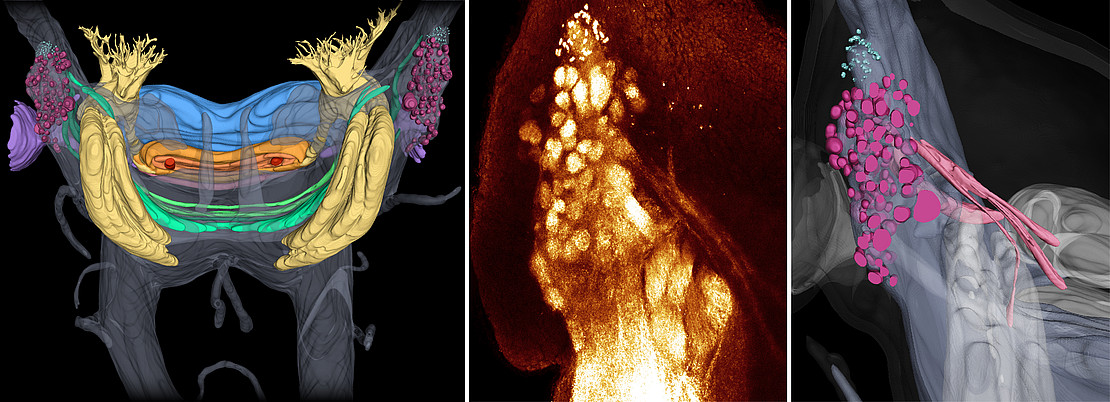 Image: Christine Martin.
Stump-footed animals (onychophores) live in tropical and temperate forests in the southern hemisphere and around the equator. The body structure of these invertebrates, most of which are about five centimeters long, has changed little since about 300 million years ago. They therefore play an important role in understanding the evolution of arthropods (spiders, centipedes, millipedes, crustaceans, and insects) the most diverse group of animals on Earth, to which the stump-footed animals are related. It is believed that the success of arthropods is related to both the segmentation of their bodies, a division into similar units or segments, and the nervous system. The study of related species can provide insight into how organ systems have evolved and changed over time.
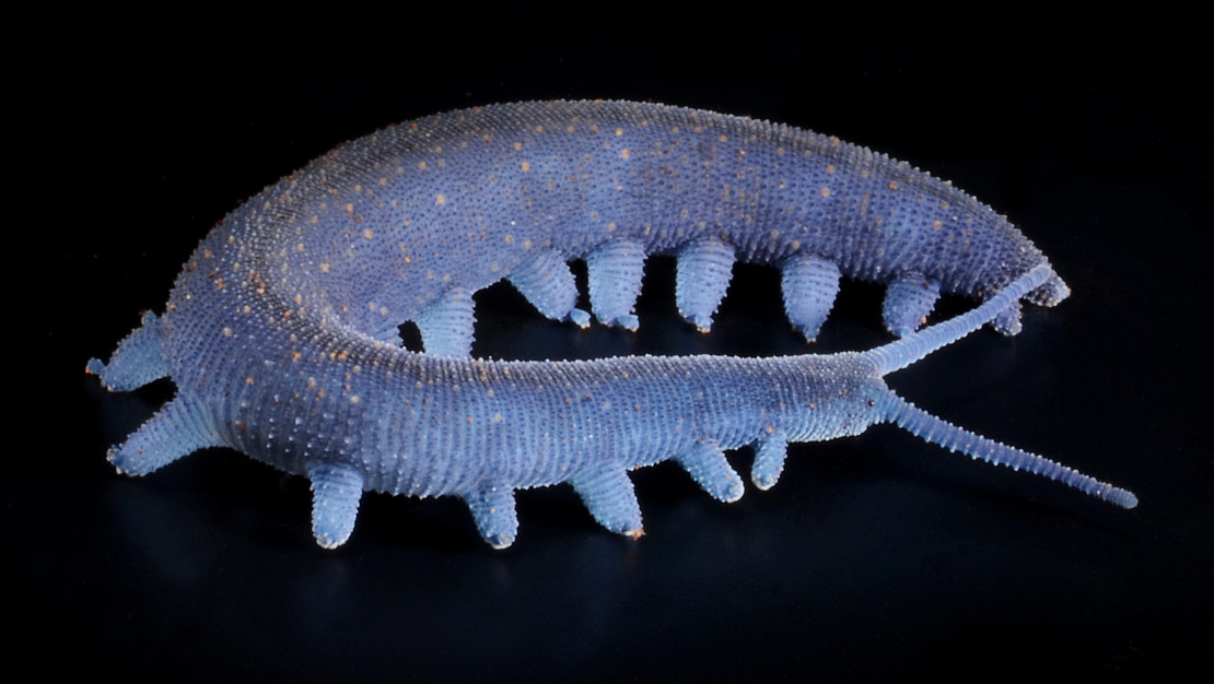 Image: Christine Martin.
"Until now, there have been inconsistencies in nomenclature and 'blind spots' on the neuroanatomical map of stump-footed animals. Our data clarify these scientific controversies and make future neuroanatomical studies of stump-footed animals more reproducible and comparable" explains Prof. Georg Mayer, head of the Department of Zoology at the University of Kassel.
Together with other zoologists and neuroscientists, he and his team combined various state-of-the-art imaging techniques for a detailed analysis of the nervous system of the stump-footed species Euperipatoides rowelli: X-ray micro-computed tomography at the German Electron Synchrotron DESY, histology, immunohistochemistry and high-resolution confocal microscopy. From these, they developed a comprehensive neuroanatomical glossary and reconstructed a three-dimensional model of the nervous system. "Because without such a model and clear terminology, comparative analyses are hardly possible" explains the study's first author Christine Martin, who is about to complete her PhD.
These analyses provide new details on already known brain areas. They thus confirm the thesis that the so-called central body and the mushroom bodies of stump-footed and arthropods can be traced back to neuroanatomical structures that already existed in their last common ancestor (homologies). In contrast, the nerve cords in the antennae and the area in the brain that serves to process odors (olfactory lobes) most likely evolved independently in stump-footed and arthropods. In contrast to previous reports, the researchers found no evidence for a second-order visual neuropil (nerve fiber plexus). They were also able to invalidate the assumption of a potential nerve node, the so-called "frontal ganglion," in the frontal region of the stub-footed head. In addition, the data revealed unprecedented details of the stump-footed brain: each mushroom body of E. rowelli consists of four lobes, not just three. The supposed "accessory peduncles" are not parts of the mushroom bodies, but independent neural connections associated with the antennal and subantennal neuropils, linking both halves of the brain. The central body of stumpy feet is more complex than previously thought. Furthermore, the authors have described other neuropil regions of unknown function, such as the frontal body and the tiny microglomeruli.
Publication:
https://bmcbiol.biomedcentral.com/articles/10.1186/s12915-021-01196-w
Contact:
Prof. Dr. Georg Mayer
Institute of Biology
Head of Department Zoology
Phone: 0561 804-4805
Email: gmayer[at]onychophora[dot]com
Press contact:
Sebastian Mense
University of Kassel
Communications, Press and Public Relations
Phone: 0561 804-1961
E-mail: presse[at]uni-kassel[dot]de