Meldung
Laser detects cancerous tissue
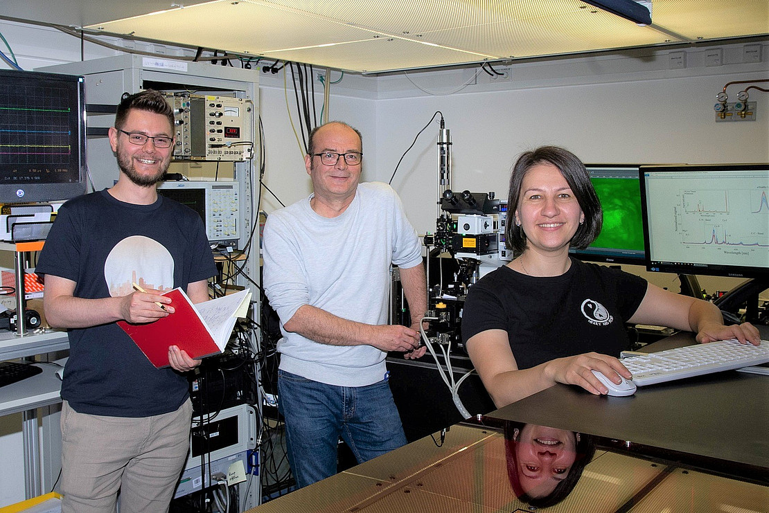 Image: Uni Kassel.
The most important method for removing cancer at an early stage is excision. However, to ensure that the tumor has been completely removed, the correct safety margin is crucial, i.e. a minimal envelope of healthy tissue must be removed around the affected tissue. A safety margin that is too small can lead to recurrence, while one that is too large can restrict the function of the affected organ.
To determine whether the malignant tissue has been completely removed, a so-called frozen section examination is often carried out. A laboratory doctor examines the removed tissue during the operation by shock freezing, cutting and staining it outside the operating theater. This allows him to determine whether the correct safety distance was maintained during removal. The further course of the operation depends on the result of this time-consuming process.
It would be desirable to have an alternative or supplementary technique with which the type of tissue operated on can be determined quickly and precisely in order to reduce the operation time and the stress on the patient.
This is where the work of the Kassel researchers comes in. Using liver cancer and breast cancer samples from the archives of the Institute of Pathology North Hesse, they achieved an accuracy of 95 to almost 100 percent in distinguishing healthy from diseased tissue using a laser method. To do this, ultra-short laser flashes lasting a few billionths of a second are sent at the tissue, causing only slight ablation of the tissue. This produces light that indicates the chemical composition of the tissue.
This process was first demonstrated on plant tissue at the Nanostructure Center at the University of Kassel twenty years ago and has now been applied to this problem. To differentiate between healthy and diseased tissue, the scientists used evaluation methods based on machine learning. The new method was developed by the Kassel experimental physicists Prof. Dr. Thomas Baumert, Arne Senftleben, Cristian Sarpe, Elena Ramela Ciobotea, Christoph Burghard Morscher, Bastian Zielinskiand Hendrike Braun in cooperation with the physician Prof. Dr. Josef Rüschoff (Institute of Pathology North Hesse).
The researchers assume that this method for rapid tissue determination will find its way into the operating theater after further research and development work. If ultrashort pulse lasers are used as cutting tools during surgery, this method can even distinguish healthy from diseased tissue directly during the incision.
Baumert: "This procedure cannot cure cancer. But it can make treatment faster, safer and gentler."
The work has been published in Nature Scientific Reports under:
"Identification of Tumor Tissue in Thin Pathological Samples via Femtosecond Laser-Induced Breakdown Spectroscopy and Machine Learning"
DOI: 10.1038/s41598-023-36155-8
Contact:
Prof. Dr. Thomas Baumert/Dr. Arne Senftleben
University of Kassel
Department of Femtosecond Spectroscopy
https://www.uni-kassel.de/fb10/institute/physik/forschungsgruppen/femtosekundenspektroskopie-und-ultraschnelle-laserkontrolle/personen/mitarbeitende
Press contact:
Sebastian Mense
University of Kassel
Communications, Press and Public Relations
Phone: +49 561 804-1961
E-mail: presse[at]uni-kassel[dot]de
www.uni-kassel.de